These glands secrete into the pectinate line, at the limit between the squamous and columnar epithelium, in the anal canal.
Blockage of these glands causes inflammation, swelling and finally development of a perianal abscess.
The abscess, which is announced by pain, redness and swelling, develops usually on the anal skin. Automatic opening of the abscess or surgical drainage results in inflammation relief, but the connection (tunnel) with the intestine remains.
This connection, which is basically the chronic stage of the abscess, is what forms an anal fistula. The fistula then looks like a small hole, from which pus oozes from time to time.
Basic principles with regard to the treatment
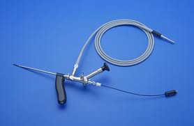
Special fistuloscope
1. Find and eliminate the internal opening.
2. Locate the tract of the fistula, and then completely excise the tract or eliminate the epithelium.
3. Make sure that the incision of the sphincters is as small as possible.
The endoscopic Video-Assisted Anal Fistula Treatment (VAAFT) is the new revolutionary treatment method for anal fistulae. For the past ten years, this method has been used in the largest medical centres around the world with great success and we are the first to introduce it in Greece.
After the examination, the specialized surgeon determines if the method can be applied to the patient, similarly to all surgery methods and techniques.
The endoscopic treatment method of anal fistulae
• Is bloodless
• Painless
• Does not require imaging examination (MRI or ultrasound)
• Does not require hospitalization
• Allows for immediate return to work
• Provides a permanent solution to the problem
• Does not require post-operative pain medication
• Presents no risk of incontinence whatsoever
The surgery includes two phases: The diagnostic and the operative one.
During the diagnostic phase, the exact fistula pathway is localized, namely the damage is traced. A special fistuloscope (an equivalent of a laparoscope) is inserted in the fistula, with irrigation running in parallel. With the fistuloscope, we trace the branches while monitoring the progress via 3D imaging on a HD screen. Tissue blockages in the tract are easily removed with a 2 mm forceps.
Then the operative phase follows, whose purpose is to destroy the fistula tract and its branches from the inside. The tract is meticulously cleaned, and all waste material is removed via suction. The epithelium of the fistula tract is then eliminated via electrocoagulation, and the fistuloscope is withdrawn. At the same time, meticulous haemostasis is performed using radiofrequencies (RF). The residual tract remains open to enable drainage of the produced secretions.
There are multiple advantages over the classic method, since no trauma is created, no dressing change is required and there is no postoperative pain. Furthermore, the perfect, enlarged imaging on a 3D HD screen allows for the execution of the surgery with high precision and unique accuracy.

Elimination of the fistula epithelium – Endoscopic image of the fistula
The new endoscopic method package includes
• Endoscope (fistuloscope) with a light source cable, a cold light source and a 3D HD screen
• Unipolar diathermy electrode or RF
• High frequency diathermy unit
• Brush
• Forceps
• Thin catheter
• Irrigation solution (5 lt. of glycine 1% and mannitol 1%)
The endoscope has an operative length of 14 cm and three channels, one for the optical source, one for suction and one for working tools insertion.


Who is Dr. Anastasios Xiarchos?

What are the days and hours that I contact him?

Which funds and insurances are supported?
