BENIGN CONDITIONS
Approximately one in five women show signs of a benign breast condition at some point in her life. 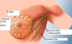 Most breast palpable masses “lumps” (8 out of 10) are benign. It might be a cyst containing fluid or a fibroadenoma (excessive growth of fibrous tissue), and can be treated easily.
If you, by chance, can feel a mass during your self-examination, you must necessarily consult a doctor, since you are not able to understand if it is benign or cancerous.
Cysts
A breast cyst is a local effusion. The cyst has smooth texture, it can be moved easily, it can be soft and most of the time it is painful. The cysts usually develop on both breasts and can develop at any site. Approximately 10% of women (1 out of 10) develop recurrent cysts (i.e. they reappear).
The cysts need hormones to develop, and that is why they are more frequent in women between 30 and 50 years old, and usually disappear after menopause. Hormone replacement therapy can trigger their initial growth or their reappearance as they were before menopause.
Fibroadenomas
They are small size lumps consisting of fibrous and glandular tissue, that cause discomfort and may grow with time or during pregnancy; that is why their removal is suggested before pregnancy. Fibroadenomas are the most common case of tumour in women of ages 20 – 30 years old, although we know they can develop at any age.
Fibroadenomas often have high mobility and can be moved easily under the skin by applying finger pressure. Once a tumour has been confirmed as a fibroadenoma, there is no need to be removed unless the patient wants so. If a tumour is less than one centimetre, it doesn’t grow or it is not painful, or if a patient has passed the age of 30, a doctor may suggest its removal.
The surgery is simple, and usually does not require hospitalization. The tumour is sent to the laboratory for microscopic examination. The removal of a fibroadenoma does not, in any way, damage the breast and does not affect the ability to breastfeed.
Fibroadenomas do not reappear if they are completely removed, but it is possible for another to develop in the future, on either breast.
Fibrocystic disease
The fibrocystic lesions of the breast are the most common cause of concern for women between the ages of 40 – 50 years, with a percentage of approximately 40% (4 out of 10). This condition can cause breast pain. The fibrocystic disease is also called fibrocystic breasts or fibrocystic breast changes. The fibrocystic lesions are caused by changes in the levels of the female hormones, oestrogens and progesterone during menstruation.
The hormones can cause dilation of blood vessels, swelling of the lactic glands and filtration of the breast pores due to breast fluid retention. It is likely that your breasts feel swollen, sore, sensitive and appear to have “lumps”.
After some time, the breasts usually return to normal, i.e. back in their previous state. However, some areas of the breast can remain harder than others, and cysts can occur in clogged or enlarged milk ducts. These areas appear to have thicker and uniform shape, and a surface full of lumps.
The fibrocystic changes can occur in both breasts, often on the upper outer quadrant where most of the mammary gland is or at the lower part of the breast. The discomfort can range from mild pain to extensive tenderness to the touch or flushing.
These changes usually, disappear after menopause, thus confirming that their cause is hormones. Fibrocystic disease treatment Not all women who develop mammary tumours have fibrocystic disease. Usually. special treatment is not necessary. The pain is generally due to hormonal swelling and will pass when the swelling subsides. Only your doctor will be able to guide you properly.
Breast cancer
Breast cancer is the most common form of cancer in women today. It rarely develops before 30 years of  age. It most commonly affects women between the ages of 50 and 70. Today, women are not usually required to undergo mastectomy (total removal of the breast), because tumour removal followed by radiation therapy can be as effective.
Risk factors: The causes of breast cancer are not yet known. It is known, however, that some situations that are called risk factors are associated with the disease. These factors are divided into three categories: A) factors we cannot change, B) factors deriving from lifestyle, C) uncertain factors.
The factors we cannot change are: gender, age, family history (breast cancer is more common in women whose mother, sister, or grandmother had suffered from the disease), race (most often occurring in the Caucasian race), previous radiation therapy, genetic factors (gene mutation).
The factors deriving from lifestyle are: childlessness (women without children or that have given birth to their first child after the age of 30, are more likely to develop breast cancer), contraceptive pills (reduce the likelihood of breast cancer), hormone replacement therapy (oestrogens with progesterone slightly raise the possibility of breast cancer), pregnancy and breastfeeding (slightly reduce the likelihood of breast cancer), as well as diet and alcohol (overweight women are more likely to develop breast cancer).
The uncertain factors are considered to be the environmental pollution from insecticides and agricultural pesticides, and smocking, especially if the smoker starts smoking in their teens.
The sooner the cancer is diagnosed, the better, as it is more likely to be successfully treated. At younger ages (20-40), a monthly self-examination is recommended. After the age of 40, women also need a clinical examination by a doctor, and a series of preventive examinations (mammography, breast ultrasound).
Breast self-examination
Visual inspection: We inspect the breasts for changes in their shape and size, changes in the shape 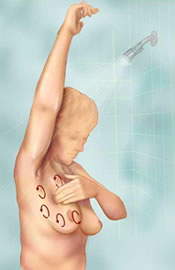 and size of the nipples, and if there are secretions from them. The examination is done in 3 ways. Standing with your hand hanging loosely down, raise the hands high above your head, observing how the breasts follow the motion and watch for any changes on the surface of the breast skin. With your hands around your waist, look for changes in the breasts contour or any “pullings” in any area.
Palpation of the breast and armpit: It is considered to be the most important examination of the breasts. The palpation can be performed while standing or lying down, and always in the same way.
Clinical examination: Women between 20-40 years of age should be clinically examined every 3 years by a doctor. Then, after 40, they should do so annually. The proper course is to have the examination before any other test (mammography, ultrasound).
Mammography: Mammography is a breast x-ray with a special machine, the mammography unit. With mammography, we get information for palpable lumps, but also for impalpable lesions. The first mammogram should be done at the age of 35-40 and should be used as reference for the future. After the age of 40, a woman should have a mammogram once a year as a preventive measure, so as to discover the potentially developed cancer at an early stage.
Breast ultrasound: It is a very simple examination that uses sound waves to form the image of the  breast. It is very sensitive, and can distinguish, for example, a cyst from a solid tumour.
Additional tests: If during the mammography and the breast ultrasound a tumour is found, additional tests are done (Magnetic Resonance Imaging, cytologic examination and biopsy).
Magnetic Resonance Imaging: Magnetic Resonance Imaging using an intravenous administration of contrast agent is the most sensitive, complementary imaging method. It is used when we want to determine the areal extent of the malignant lesion, in case of invasive lobular breast cancer, and preoperatively. In no case should Magnetic Resonance Imaging substitute classic mammography and ultrasounds. It is a complementary method. No examination can substitute another, one complements the other to check for cancer spreading to other parts of the body.
TREATMENT
Today, the weapons we have against breast cancer can be divided into 2 categories.
1. Local treatment aims at treating the tumour without affecting the rest of the body, and involves surgical removal and radiotherapy.
2. Systemic therapy, during which we intravenously or orally administer medicine to treat cancer cells that have spread outside the breast, and involves hormone therapy and chemotherapy.
LIFE AFTER DIAGNOSIS
The diagnosis of breast cancer is a very traumatic experience for most women. Many women feel frustrated and anxious. They are thinking about the changes that they have to make to their lives, because they need to be hospitalized, and to undergo treatments.
The treatment can cause tiredness, lethargy and poor psychological condition, that they will quickly overcome. In case you feel that way, talk to your partner or other people close to you, even with women who have the same condition. Many women become confident this way.
Chemotherapy and radiation treatment do not affect the sexuality of women. Finally, we must not forget that many women each day face the harsh reality of breast cancer. It is good talking to them, and collectively face fear, anxiety and problems.